
Keywords
MRI
Radiculopathy
LS Spine
How to Cite
Abstract
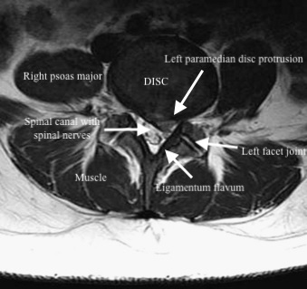
Introduction: Chronic low back pain (LBP) is a common cause of disability worldwide. Magnetic resonance imaging (MRI) is an excellent non-invasive imaging modality for morphologic evaluation of the lumbar spine in patients with chronic low back pain because of its high contrast resolution and lack of ionizing radiation. This study was done to see the patterns of MRI changes in patients with chronic low back pain in a tertiary care center in Western Nepal. Methods: This was a cross-sectional study conducted on patients presenting with chronic low back pain. Eleven MRI parameters were noted and analyzed. Chi square test and Fisher’s Exact test were employed to see the associations between the various MRI findings. Results: A total of 108 patients were evaluated during the study period. MRI changes were noted in over 95% of the cases. Degenerative changes were the most common cause of low back pain, disc bulge being the most common MRI finding. A significant association was found between radiculopathy and decreased lumbar lordosis and vertebral endplate changes. Conclusion: MRI is an invaluable tool in the evaluation of chronic LBP because of its high resolution and lack of ionizing radiation. Significant MRI findings are noted in most of the cases of chronic LBP, degenerative changes being the most common and ranging from congenital to malignant lesions.
References
Chou R. Low back pain (chronic). BMJ clinical evidence. 2010 Oct 8;2010:1116.PMID: 21418678 [Publisher Full Text]
Hoy D, March L, Brooks P, Blyth F, Woolf A, Bain C, et al. The global burden of low back pain: estimates from the Global Burden of Disease 2010 study. Annals of the Rheumatic Diseases. 2014 Jun 1;73(6):968–74. PMID: 24665116 doi:10.1136/annrheumdis-2013-204428
Deyo RA, Weinstein JN. Low back pain. New England Journal of Medicine. 2001 Feb 1;344(5):363–70. PMID: 11172169 DOI: 10.1056/NEJM200102013440508
Suthar P, Patel R, Mehta C, Patel N. MRI evaluation of lumbar disc degenerative disease. Journal of Clinical and Diagnostic Research. 2015 Apr;9(4):TC04-09. PMID: 26023617 DOI: 10.7860/JCDR/2015/11927.5761 [Publisher Full Text]
Hong SH, Choi J-Y, Lee JW, Kim NR, Choi J-A, Kang HS. MR imaging assessment of the spine: infection or an imitation? Radiographics. 2009 Apr;29(2):599–612. PMID:19325068 DOI: 10.1148/rg.292085137
Modic MT, Ross JS. Lumbar degenerative disk disease. Radiology. 2007 Oct;245(1):43–61. PMID: 17885180 DOI: 10.1148/radiol.2451051706
Okpala F. Measurement of Lumbosacral Angle in Normal Radiographs: A Retrospective Study in Southeast Nigeria. Annals of Medical Health and Science Research. 2014;4(5):757–62. PMID: 25328789 DOI:10.4103/2141-9248.141548 [Publisher Full Text]
Been E, Kalichman L. Lumbar lordosis. The Spine Journal. 2014 Jan;14(1):87–97. PMID: 24095099 DOI: https://doi. org/10.1016/j.spinee.2013.07.464
Modic MT, Steinberg PM, Ross JS, Masaryk TJ, Carter JR. Degenerative disk disease: assessment of changes in vertebral body marrow with MR imaging. Radiology. 1988 Jan 1;166(1):193–9. PMID:3336678 DOI: 10.1148/ radiology.166.1.3336678
Gopalakrishnan N, Nadhamuni K, Karthikeyan T. Categorization of Pathology Causing Low Back Pain using Magnetic Resonance Imaging (MRI). Journal of Clinical and Diagnostic Research. 2015 Jan;9(1):TC17-20. PMID: 25738056 DOI: 10.7860/JCDR/2015/10951.5470 [Publisher Full Text]
Biluts H, Munie T, Abebe M. Review of lumbar disc diseases at TikurAnbessa Hospital. Ethiopian Medical Journal. 2012 Jan;50(1):57–65. PMID: 22519162
Dahal S, Joshi A, Pant S. Spectrum of Lumbar Spine Pathologies in Patients with Low Back Pain on MR Examination: A Retrospective Hospital Based Study. Post-Graduate Medical Journal of NAMS. 2015;12(02). [Publisher Full Text]
Ansari MA, Subedi K, Panta OB, Suwal S. MRI pattern of lumbosacral degeneration in Tribhuvan University Teaching Hospital, Nepal. Journal of institute of Medicine. 2015;38(2). [Publisher Full Text]
Ogon I, Takebayashi T, Takashima H, Tanimoto K, Ida K, Yoshimoto M, et al. Analysis of chronic low back pain with magnetic resonance imaging T2 mapping of lumbar intervertebral disc. Journal of Orthopaedic Science. 2015 Mar 1;20(2):295–301. PMID: 25649736 DOI: 10.1007/ s00776-014-0686-0
Nardin RA, Patel MR, Gudas TF, Rutkove SB, Raynor EM. Electromyography and magnetic resonance imaging in the evaluation of radiculopathy. Muscle Nerve. 1999 Feb;22(2):151–5.PMID: 10024127
Sakamaki T, Sairyo K, Sakai T, Tamura T, Okada Y, Mikami H. Measurements of ligamentum flavum thickening at lumbar spine using MRI. Archives of Orthopaedic Trauma and Surgery. 2009 Oct;129(10):1415–9. PMID: 19280205 DOI:10.1007/s00402-009-0849-1
Saleem S, Aslam HM, Rehmani MAK, Raees A, Alvi AA, Ashraf J. Lumbar disc degenerative disease: disc degeneration symptoms and magnetic resonance image findings. Asian Spine Journal. 2013 Dec;7(4):322–34. PMID: 24353850 DOI: 10.4184/asj.2013.7.4.322 [Publisher Full Text]
Roudsari B, Jarvik JG. Lumbar Spine MRI for Low Back Pain: Indications and Yield. American Journal of Roentgenology. 2010 Sep 1;195(3):550–9. PMID: 20729428 doi:10.2214/AJR.10.4367
- The Journal of Lumbini Medical College (JLMC) publishes open access articles under the terms of the Creative Commons Attribution (CC BY) License which permits use, distribution and reproduction in any medium, provided the original work is properly cited.
- JLMC requires an exclusive license to publish the article first in its journal in print and online.
- The corresponding author should read and agree to the following statement before submission of the manuscript for publication,
- License agreement
- In submitting an article to Journal of Lumbini Medical College (JLMC) I certify that:
- I am authorized by my co-authors to enter into these arrangements.
- I warrant, on behalf of myself and my co-authors, that:
- the article is original, has not been formally published in any other peer-reviewed journal, is not under consideration by any other journal and does not infringe any existing copyright or any other third-party rights;
- I am/we are the sole author(s) of the article and have full authority to enter into this agreement and in granting rights to JLMC are not in breach of any other obligation;
- the article contains nothing that is unlawful, libelous, or which would, if published, constitute a breach of contract or of confidence or of commitment given to secrecy;
- I/we have taken due care to ensure the integrity of the article. To my/our - and currently accepted scientific - knowledge all statements contained in it purporting to be facts are true and any formula or instruction contained in the article will not, if followed accurately, cause any injury, illness or damage to the user.
- I, and all co-authors, agree that the article, if editorially accepted for publication, shall be licensed under the Creative Commons Attribution License 4.0. If the law requires that the article be published in the public domain, I/we will notify JLMC at the time of submission, and in such cases the article shall be released under the Creative Commons 1.0 Public Domain Dedication waiver. For the avoidance of doubt it is stated that sections 1 and 2 of this license agreement shall apply and prevail regardless of whether the article is published under Creative Commons Attribution License 4.0 or the Creative Commons 1.0 Public Domain Dedication waiver.
- I, and all co-authors, agree that, if the article is editorially accepted for publication in JLMC, data included in the article shall be made available under the Creative Commons 1.0 Public Domain Dedication waiver, unless otherwise stated. For the avoidance of doubt it is stated that sections 1, 2, and 3 of this license agreement shall apply and prevail.
Please visit Creative Commons web page for details of the terms.