
Keywords
Computed tomography
Portal vein
How to Cite
Abstract
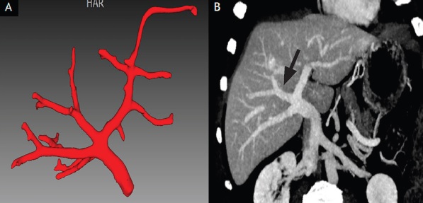
Introduction: Various anatomical variants are encountered in portal venous system which are quite important while undergoing hepatobiliary surgeries and percutaneous radiological interventions. Contrast enhanced computed tomography (CECT) of the abdomen is considered a better imaging modality to identify these variations. Methods: A descriptive prospective study was conducted in 1000 individuals undergoing CECT of abdomen. Triple phase CECT scan of the abdomen was done and the portal vein anatomy was reconstructed and analyzed. Results: Normal branching pattern of the portal vein was seen in 786 (78.6%) patients. Variations were seen in rest of the 214 (21.40%) patients. The most common variant was trifurcation of the portal vein seen in 113 (11.3%) patients. Right posterior portal vein as the first branch of main portal portal vein was found in 72 (7.2%) patients. Right anterior portal vein arising from left portal vein was seen in 29 (2.9%). Sixty nine of the 567 males had trifurcation accounting for 12.1% incidence of this variation amongst males. Trifurcation was seen in 44 of the 433 females resulting in an incidence of 10.1%. Forty-four (7.7%) males and 28 (6.4%) females had right posterior portal vein as the first branch of main portal vein. Right anterior portal vein was noted to arise from the left portal vein in 20 (3.5%) males and nine (2.07%) females. Conclusion: The most common variation in portal venous system was trifurcation of portal vein followed by right posterior as first branch and right anterior branch arising from left portal vein respectively.
References
Carneiro C, Brito J, Bilreiro C, Barros M, Bahia C, Santiago I, et al. All about portal vein: a pictorial display to anatomy, variants and physiopathology. Insights Imaging. 2019;10(1):38. PMID: 30900187 DOI: https://doi.org/10.1186/s13244-019-0716-8
Corness JAG, McHugh K, Roebuck DJ, Taylor AM. The portal vein in children: radiological review of congenital anomalies and acquired abnormalities. Pediatr Radiol. 2006;36(2):87- 96. PMID: 16284764 DOI: https://pubmed.ncbi.nlm.nih.gov/16284764/
Strasberg S. Hepatic, biliary and pancreatic anatomy. In: Garden JO, Parks RW (eds). Hepatobiliary and Pancreatic Surgery. 5th ed. UK: Elsevier; 2013. p.17-38.
Sureka B, Patidar Y, Bansal K, Rajesh S, Agrawal N, Arora A. Portal vein variations in 1000 patients: surgical and radiological importance. British Journal of Radiology. 2015;88(1055):20150326. DOI: https://doi.org/10.1259/bjr.20150326
Lee WK, Chang SD, Duddalwar VA, Comin JM, Perera W, Lau WFE, et al. Imaging assessment of congenital and acquired abnormalities of the portal venous system. Radiographics. 2011;31(4):905-26. PMID: 21768231 DOI: https://doi.org/10.1148/rg.314105104
Dighe M, Vaidya S. Case report. Duplication of the portal vein: a rare congenital anomaly. Br J Radiol. 2009;82(974):e32-4. PMID: 19168687 DOI: https://doi.org/10.1259/bjr/81921288
Guerra A, De Gaetano AM, Infante A, Mele C, Marini MG, Rinninella E, et al. Imaging assessment of portal venous system: pictorial essay of normal anatomy, anatomic variants and congenital anomalies. Eur Rev Med Pharmacol Sci. 2017;21(20):4477-4486. PMID: 29131270.
Covey AM, Brody LA, Getrajdman GI, Sofocleous CT, Brown KT. Incidence, patterns, and clinical relevance of variant portal vein anatomy. American Journal of Roentgenology. 2004;183(4):1055-64. DOI: https://www.ajronline.org/doi/10.2214/ajr.183.4.1831055
Atri M, Bret PM, Fraser-Hill MA. Intrahepatic portal venous variations: prevalence with US. Radiology. 1992;184(1):157-8. PMID: 1609075 DOI: https://doi.org/10.1148/radiology.184.1.1609075
Fraser-Hill MA, Atri M, Bret PM, Aldis AE, Illescas FF, Herschorn SD. Intrahepatic portal venous system: variations demonstrated with duplex and color Doppler US. Radiology. 1990;177(2):523-6. PMID: 2217795 DOI: https://doi.org/10.1148/radiology.177.2.2217795
Atasoy C, Ozyürek E. Prevalence and types of main and right portal vein branching variations on MDCT. AJR Am J Roentgenol. 2006;187(3):676- 81. PMID: 16928929 DOI: https://pubmed.ncbi.nlm.nih.gov/16928929/
- The Journal of Lumbini Medical College (JLMC) publishes open access articles under the terms of the Creative Commons Attribution (CC BY) License which permits use, distribution and reproduction in any medium, provided the original work is properly cited.
- JLMC requires an exclusive license to publish the article first in its journal in print and online.
- The corresponding author should read and agree to the following statement before submission of the manuscript for publication,
- License agreement
- In submitting an article to Journal of Lumbini Medical College (JLMC) I certify that:
- I am authorized by my co-authors to enter into these arrangements.
- I warrant, on behalf of myself and my co-authors, that:
- the article is original, has not been formally published in any other peer-reviewed journal, is not under consideration by any other journal and does not infringe any existing copyright or any other third-party rights;
- I am/we are the sole author(s) of the article and have full authority to enter into this agreement and in granting rights to JLMC are not in breach of any other obligation;
- the article contains nothing that is unlawful, libelous, or which would, if published, constitute a breach of contract or of confidence or of commitment given to secrecy;
- I/we have taken due care to ensure the integrity of the article. To my/our - and currently accepted scientific - knowledge all statements contained in it purporting to be facts are true and any formula or instruction contained in the article will not, if followed accurately, cause any injury, illness or damage to the user.
- I, and all co-authors, agree that the article, if editorially accepted for publication, shall be licensed under the Creative Commons Attribution License 4.0. If the law requires that the article be published in the public domain, I/we will notify JLMC at the time of submission, and in such cases the article shall be released under the Creative Commons 1.0 Public Domain Dedication waiver. For the avoidance of doubt it is stated that sections 1 and 2 of this license agreement shall apply and prevail regardless of whether the article is published under Creative Commons Attribution License 4.0 or the Creative Commons 1.0 Public Domain Dedication waiver.
- I, and all co-authors, agree that, if the article is editorially accepted for publication in JLMC, data included in the article shall be made available under the Creative Commons 1.0 Public Domain Dedication waiver, unless otherwise stated. For the avoidance of doubt it is stated that sections 1, 2, and 3 of this license agreement shall apply and prevail.
Please visit Creative Commons web page for details of the terms.