
Keywords
Fundus fluorescence angiography
Optical coherence tomography
How to Cite
Abstract
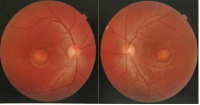
Introduction: Best disease or vitelliform macular dystrophy is a rare autosomal dominant disorder with bilateral presentation characterized by subretinal accumulation of yellowish material in the macular area. Macular findings range from a small yellow spot, multiple vitelliform or atrophic lesions to a chorio-retinal scar. Case report: A 35 years male presented to our outpatient department with chief complaint of blurring of vision of both eyes for the past three months. His visual acuity was 6/12 in both right and left eyes. On slit-lamp bio-examination anterior segments of both eyes were normal. On fundus examination, both eyes revealed a single, circular, yellow-opaque egg yolk-like macular lesion with no other abnormalities. Optical coherence tomography of both eyes revealed deposits of homogenous hyper- reflective material beneath retinal pigment epithelium at fovea. Fundus fluorescence angiography showed blocked fluorescence at the site of vitelliform lesion of both eyes. Conclusion: Best vitelliform macular dystrophy is a rare genetic disorder with incomplete penetrance. Optical coherence tomography and fundus fluorescence angiography support the diagnosis.
References
Deutman AF, Hoyng CB. Macular Dystrophies. In: Schachat AP, Hengst TC. (eds.) Medical Retina. 3rd ed. London: Mosby; 2001. p.1225
Querques G, Zerbib J, Santacroce R, Margaglione M, Delphin N, Rozet JM, et al. Functional and clinical data of Best vitelliform macular dystrophy patients with mutations in the BEST1 gene. Mol Vis. 2009;15:2960-72. PMID: 20057903
Stone EM, Nichols BE, Sterb LM, Kimura AE, Sheffield VC. Genetic linkage of vitelliform macular degeneration (Best’s disease) to chromosome 11q13. Nat Genet. 1992;1(4):246- 50. PMID: 1302019. DOI: https://doi.org/10.1038/ ng0792-246
Frangieh GT, Green WR, Fine SL. A histopathologic Study of Best’s Macular Dystrophy. Arch Ophthalmol. 1982;100(7):1115- 21. PMID: 7092655. DOI: https://doi.org/10.1001/ archopht.1982.01030040093017
Mohler CW, Fine SL. Long-term evaluation of patients with Best’s vitelliform dystrophy. Ophthalmology. 1981:88(7):688-92. PMID: 7267039. DOI: https://doi.org/10.1016/s0161- 6420(81)34965-3
Hartzell HC, Qu Z, Yu K, Xiao Q, Chien LT. Molecular physiology of bestrophins: multifunctional membrane proteins linked to Best disease and other retinopathies. Physiol Rev. 2008;88(2):639-72. PMID: 18391176. DOI: https://doi.org/10.1152/physrev.00022.2007
Weingeist TA, Kobrin J, Watzke RC. Histopathology of Best’s macular dystrophy. Arch Ophthalmol. 1982;100(7):1108-14. PMID: 7092654. DOI: https://doi.org/10.1001/ archopht.1982.01030040086016
Schmitz-Valckenberg S, Holz FG, Bird AC, Spaide RF. Fundus autofluorescence imaging: review and perspectives. Retina. 2008;28(3):385-409. PMID: 18327131. DOI: https://doi.org/10.1097/ IAE.0b013e318164a907
- The Journal of Lumbini Medical College (JLMC) publishes open access articles under the terms of the Creative Commons Attribution (CC BY) License which permits use, distribution and reproduction in any medium, provided the original work is properly cited.
- JLMC requires an exclusive license to publish the article first in its journal in print and online.
- The corresponding author should read and agree to the following statement before submission of the manuscript for publication,
- License agreement
- In submitting an article to Journal of Lumbini Medical College (JLMC) I certify that:
- I am authorized by my co-authors to enter into these arrangements.
- I warrant, on behalf of myself and my co-authors, that:
- the article is original, has not been formally published in any other peer-reviewed journal, is not under consideration by any other journal and does not infringe any existing copyright or any other third-party rights;
- I am/we are the sole author(s) of the article and have full authority to enter into this agreement and in granting rights to JLMC are not in breach of any other obligation;
- the article contains nothing that is unlawful, libelous, or which would, if published, constitute a breach of contract or of confidence or of commitment given to secrecy;
- I/we have taken due care to ensure the integrity of the article. To my/our - and currently accepted scientific - knowledge all statements contained in it purporting to be facts are true and any formula or instruction contained in the article will not, if followed accurately, cause any injury, illness or damage to the user.
- I, and all co-authors, agree that the article, if editorially accepted for publication, shall be licensed under the Creative Commons Attribution License 4.0. If the law requires that the article be published in the public domain, I/we will notify JLMC at the time of submission, and in such cases the article shall be released under the Creative Commons 1.0 Public Domain Dedication waiver. For the avoidance of doubt it is stated that sections 1 and 2 of this license agreement shall apply and prevail regardless of whether the article is published under Creative Commons Attribution License 4.0 or the Creative Commons 1.0 Public Domain Dedication waiver.
- I, and all co-authors, agree that, if the article is editorially accepted for publication in JLMC, data included in the article shall be made available under the Creative Commons 1.0 Public Domain Dedication waiver, unless otherwise stated. For the avoidance of doubt it is stated that sections 1, 2, and 3 of this license agreement shall apply and prevail.
Please visit Creative Commons web page for details of the terms.